
Photographs from the anatomical collection of
National Museum of Health and Medicine
Armed Forces Institute of Pathology
Washington D.C.
June 2007
&
Silver Spring, Maryland
January 2015

No. 3694
When I first visited The National Museum of Health and Medicine of the Armed Forces Institute of Pathology (NMHM/AFIP) in 2007, it was located on the campus of Walter Reed Army Medical Center in Washington D.C. The museum was originally founded as the Army Medical Museum (AMM) in 1862. The initial purpose of the institution was to document the effects of battle wounds and disease, and to study and improve medical practice. The American Civil War was underway at the time, so there was no shortage of study material to be collected. Today, NMHM (which acquired that name in 1989) maintains a collection of more than twenty-four million items. The collection includes archives, medical equipment, and human remains both normal and pathological for display and research. Perhaps the best known objects in the collection relate to the autopsies of President Abraham Lincoln (including the bullet that killed him, the probe used to find the bullet, and the surgeon’s blood-stained shirt cuff) and his assassin, John Wilkes Booth (a section of his spine).
The Armed Forces Institute of Pathology (AFIP) was decommissioned in September 2011. About the same time, the NMHM was forced to leave its home at Walter Reed due to the closure of the medical center. In 2012, the NMHM reopened in a new but smaller space at U.S. Army Garrison-Forest Glen Annex in nearby Silver Spring, Maryland. I made my first visit to the new site in 2015.
Special thanks to Brian Spatola and Tim Clarke, Jr. for their hospitality and enthusism for this project.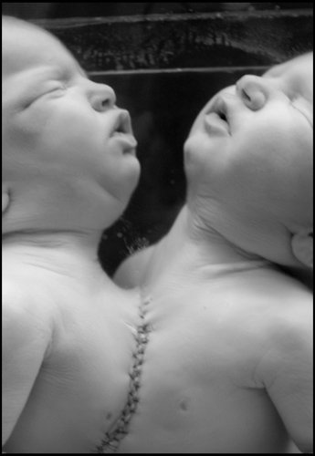
Conjoined twins

Black and white photo of a doll with a human face and painted facial features, sitting against a background.

A human anatomy model showing a cutaway view of the chest and head, with the internal organs and skeleton visible, lying on a wooden surface.
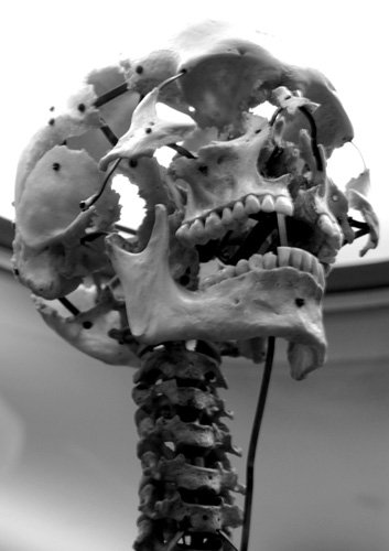
An expanded human skull, featuring a prominent jaw with teeth and a spine extending downward.

A black and white photo containing two exposures from a roll of 35mm film showing a human skeleton with its skull visible, taken from below, showing the ribcage and spinal column.

Black and white photo of a human skeleton with expanded skull visible, placed beneath a bright lighting fixture.
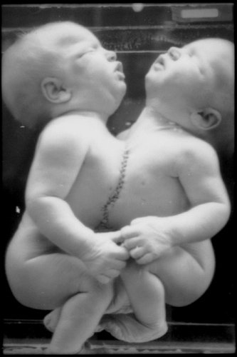
Black and white photo of conjoined twins submerged in a jar of preservative liquid on display in a museum.

A black and white photo containing two exposures from a roll of 35mm film showing preserved specimens of conjoined twins at left and a fetus with sirenomelia (a.k.a., mermaid syndrome) at right.

Close-up black and white photo of conjoined twins submerged in a jar of preservative liquid on display in a museum.

Black and white photo of an human fetus with sirenomelia ("mermaid syndrome") submerged in a jar of preservative liquid on display in a museum.

Two human skeletons facing forward in a black-and-white photo.
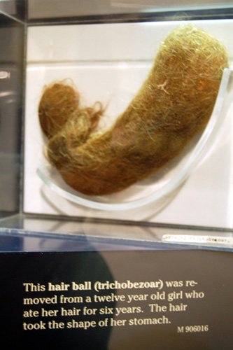
A hairball, shaped like a stomach, on display in a glass case with a caption explaining it was removed from a young girl.

Close-up view of a vintage microscope.

The Kentucky Mummy

Abnormal specimens

Sirenomelia (Mermaid syndrome)

Dwarfism

Black and white photograph of an anatomical model of a person with a distorted face, prominent lumps on the nose, and a pipe near the mouth.
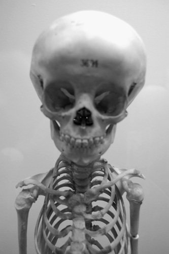
A human skeleton with a skull that has the number '36' written on it, displayed against a plain background.

No. 3765

No. 3698

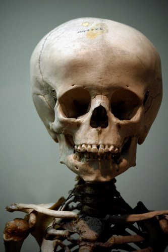
A human skeleton with a large skull in a black and white photograph.

Ossification

Musket shot
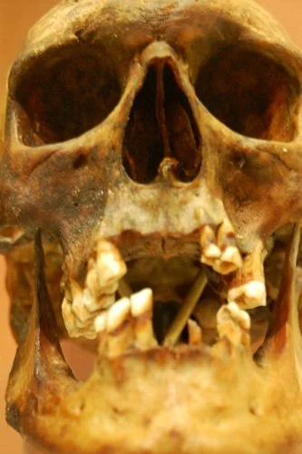
Exhumed skull

A close-up profile of a human skull highlighting an arrowhead lodged the eye socket, in black and white.

Black and white photo of a clarified human fetus in preservative liquid on display in a museum.

Anantomicla model of a human head with part of the face peeled back revealing underlying tissue and muscles, with a simialr preserved anatomical specimen in the background.

A detailed anatomical preparation of a preserved human face with muscles, bones, and tissues exposed, showing a reflection of the face from the side of the container.

Close-up of a preserved specimen of a dissected face showing muscles, bones, and exposed tissues.

Close-up image of three preserved and partially dissected human hands on display in a museum for study.

Close-up of an anatomical model of a person with a large tumor, in black and white.
502963: 5 years

A row of human skulls and skeletons displayed closely together.

Black and white photo of a human skull showing the top of the skull with writing and numbers on it.

M-906046: Anencephaly

Close-up of conjoined twins submerged in a jar of preservative liquid on display in a museum.

Preserved fetus with fists clenched, head turned to the side, in black and white.

Close-up of a clarified fetus with orange and red markings in a glass container of preservative fluid.

1989.0012.10: Cleared and stained 6-month-old human foetus
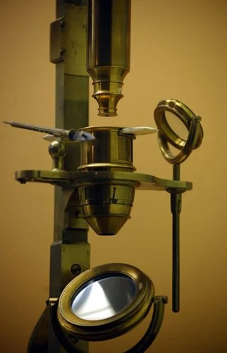
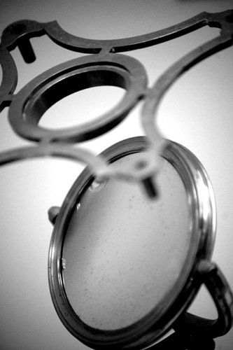
A microscope with a circular mirror used for illumination.

Diving mask and snorkel equipment

Close-up of a vintage telescope and mechanical arms in black and white.

Close-up of a clarified fetus with orange and red markings in a glass container of preservative fluid for educational and scientific study

Close-up of preserved conjoined twins, showing linked hands.

Close-up of a preserved fetus curled inside a womb in a black-and-white photograph.
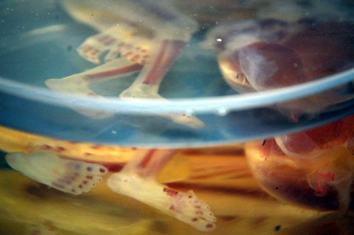
Close-up of a clarified fetus with orange and red markings in a glass container of preservative fluid with blurred reflections.

A close-up black and white image of a clarified fetus in a container of preservative fluid.

Close-up of a vintage microscope with a focusing knob and a mirror.
Close-up of a vintage microscope with a focusing knob and a mirror.

Close-up of a microscope with its eyepiece and objective lens visible.

Close-up of a vintage microscope with a focusing knob and a mirror.

Abnormal human fetus specimens submerged in preservative liquid on display in a museum.

Two newborn babies laying side by side with their heads touching, showing similar facial features.

Two human skeletons with skulls displayed in a museum exhibit.

Three skeletons displayed standing side by side

A black and white photo of a human skull atop a skeleton's chest, set against a dotted background.

Four human skeletons of varying sizes displayed in a glass case, with a spotlight overhead illuminating them.