|
|
Photographs and drawings from the anatomical collection of
The Mütter Museum,
College of Physicians of Philadelphia
April and August 2008

|
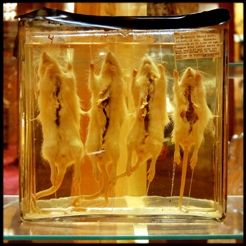
No. 15-2-11: Hypertrophied rats
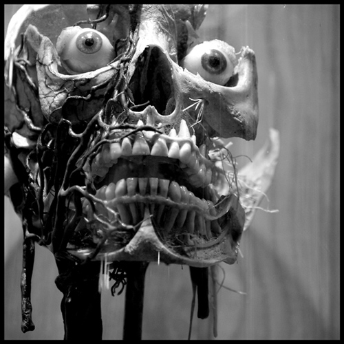
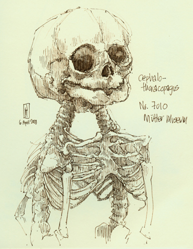
No. 7010: Cephalo-thoracopagus
ink drawing
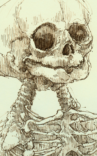
detail: No. 7010: Cephalo-thoracopagus
ink drawing
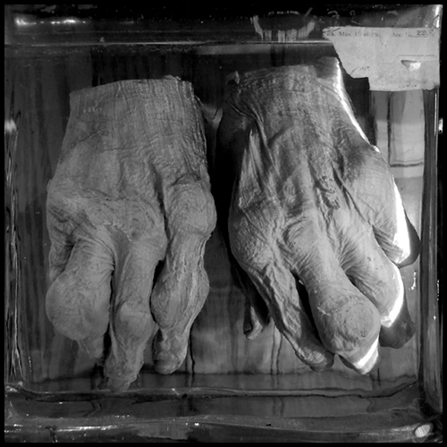
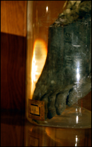
No. 2202.1: Two hands with gout
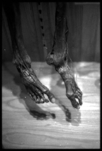
McClellan skeleton
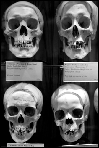
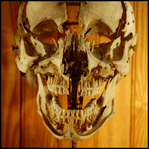
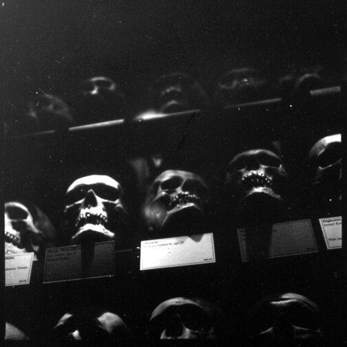
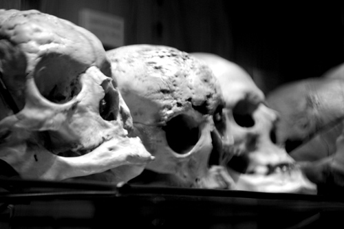
Hyrtl Skull Collection No. 1
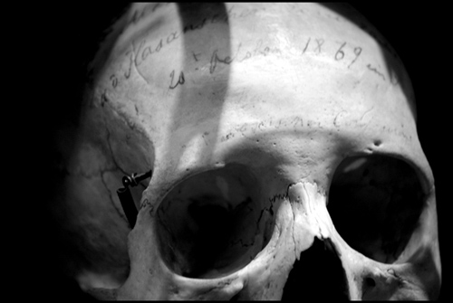
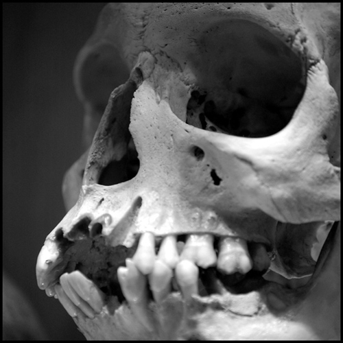
Austrian anatomist and phrenologist Joseph Hyrtl (1810-1894) amassed a large collection of human skulls and other bones from around the world for use in comparative anatomy studies. The Mütter Museum acquired more than 100 skulls from his collection in 1874.
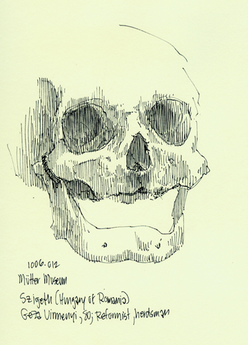
No. 1006.012: Szigeth
ink drawing
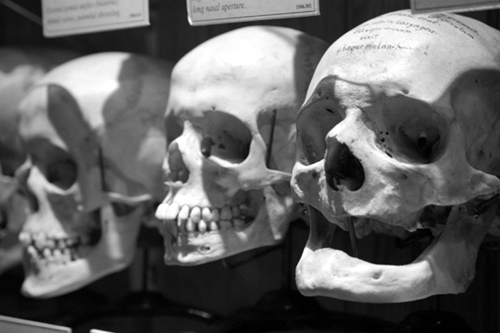
Hyrtl Skull Collection No. 3
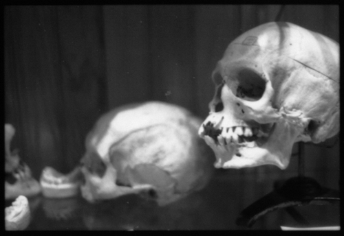
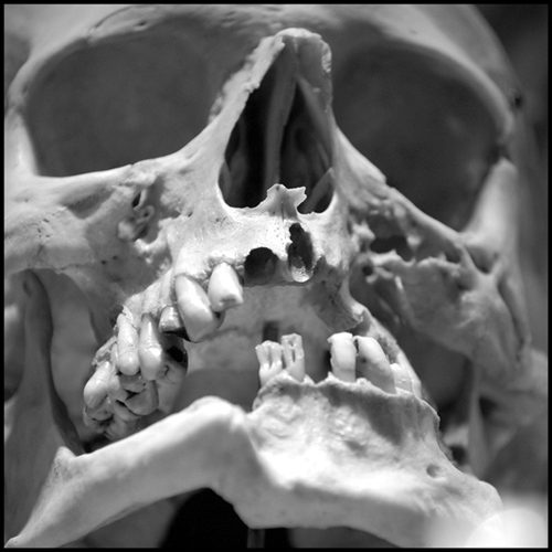
Ammended (Hyrtl Skull Collection)
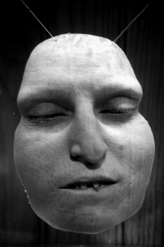
Suspended profile
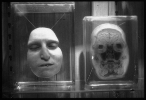
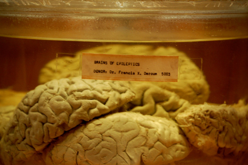
Brains of epileptics
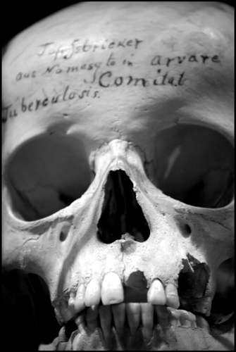
Tuberculosis (Hyrtl Skull Collection)
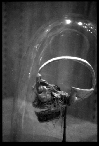
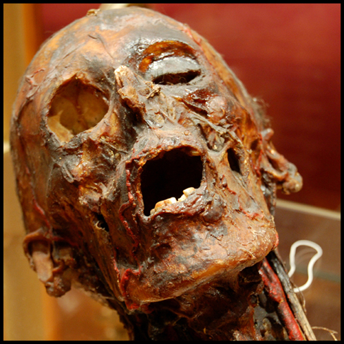
No. F1993.28A
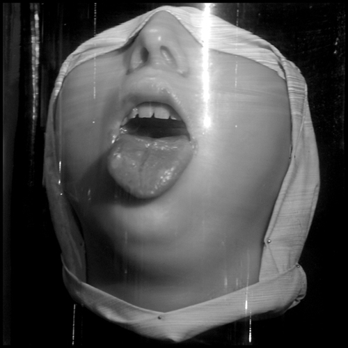
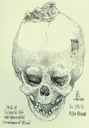
No. 1145.70
ink drawing
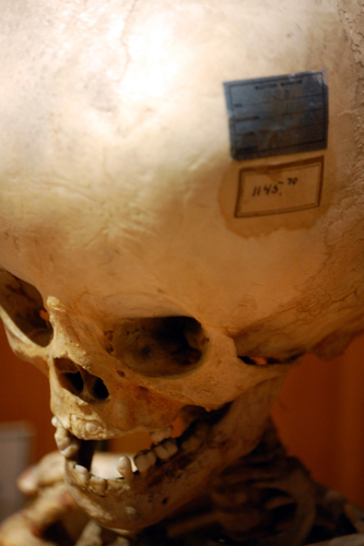
No. 1145.70 (Hydrocephalus)
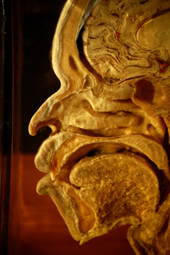
Section
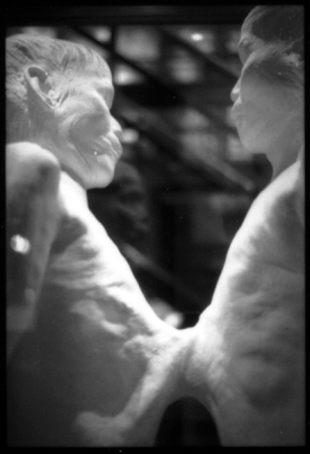
Chang & Eng No. 3
Plaster death cast of Chang & Eng Bunker (1811-1874), the original Siamese Twins. During their long career the twins
amassed and lost several fortunes. In the 1840s, Chang and Eng became naturalized American citizens and retired from show
business to live quiet lives as Southern planters. In 1843, they each married — sisters Adelaide and Sarah Yates — and began to father
numerous children, twenty-one between them. In the wake of the Civil War, the twins were forced to return to show business to recoup their financial losses.
When Chang and Eng died during the night of 16-17 January 1874 (Chang first of an apparent stroke, then Eng several hours later due to exsanguination), Drs. William Pancoast and Harrison Allen
from the College of Physicians traveled to Mount Airy, North Carolina, to collect the bodies for transportation back to Philadelphia for autopsy. As the twins had died some weeks before the doctors arrived,
it was necessary to disinter the remains and quickly embalm them. The bodies were then soldered into a tin box for transportation back to the College, where a more
thorough embalming procedure was undertaken prior to the autopsy on Monday, 9 February 1874.
During the autopsy it was discovered that the twins shared a conjoined liver, which is now displayed in a large jar beneath their death cast.
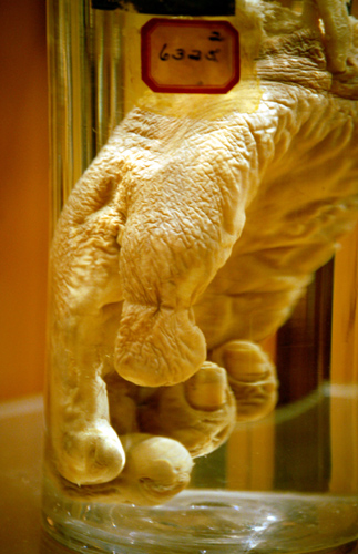
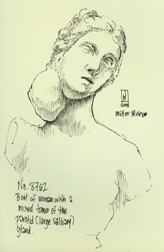
No. 8762
ink drawing
One of the more amusing trends in early 19th-century model making was the custom of taking classical sculptures and applying deformities to them,
as is the case here where an unlucky Diana or Aphrodite now finds herself with a salivary gland tumor. Another example appears in the previous photograph,
where a Christ child straight out of a nativity scene (note the hand raised in blessing) had a large tumor applied to its buttocks. This strange work of art (probably acquired in France)
is displayed in a case beside an actual specimen of a child with the same condition, bit with a far less serene expression.
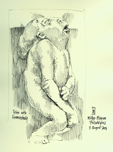
Fetus with exencephaly
ink drawing
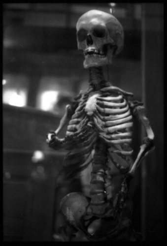
Skeleton of Mary Ashberry
Skeleton of Mary Ashberry, 3’ 6” tall, who died in 1856. Ms. Ashberry became pregnant, but tragically Mary's pelvis was too contracted to
allow the natural birth of her child and they both died in the attempt.
Hyrtl Skull Collection No. 2
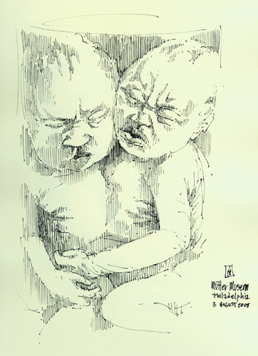
Conjoined twins
ink drawing
All drawings and photographs by James G. Mundie.
[Reproduction in any form without express written permission
of the artist is prohibited.]
|
|

|

|
All Images and Text © James G. Mundie 2008 - 2013
|

